If you have been told you might have a hernia, it is natural to wonder whether you need medical imaging to confirm it. The short answer is: not always. At The Iskandar Complex Hernia Center, Dr. Iskandar begins with a detailed history and physical examination because most clinically significant hernias can be diagnosed without additional testing. Imaging is reserved for specific situations where it truly adds value to the diagnosis and surgical plan.
Can a Hernia Be Diagnosed with a Physical Examination Alone?
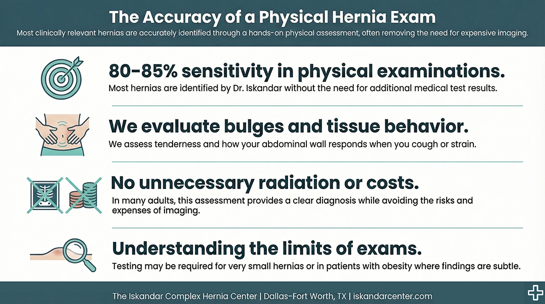
Yes, in most cases it can. The majority of clinically relevant hernias, whether ventral or inguinal, are detected through a thorough physical examination. The sensitivity of an exam is approximately 80–85%, which means most hernias are identified without the need for additional medical test results.
During the visit, Dr. Iskandar evaluates the abdomen and groin for a bulge, tenderness, and other signs and symptoms. He assesses how the tissue behaves when you cough or strain and determines whether the structure of the abdominal wall or muscle layer has been disrupted. In many adults, this hands-on assessment provides a clear medical diagnosis without exposing the patient to unnecessary radiation or cost.
However, the exam is less sensitive for very small or occult hernias and in patients with obesity, where excess tissue can make subtle findings more difficult to detect.
How Does Dr. Iskandar Decide If Imaging Is Needed?
Dr. Iskandar always examines the patient first. The physical exam guides the next steps and helps determine whether imaging is needed at all. Routine imaging for every suspected hernia is not recommended because it often adds cost and, in some cases, radiation exposure without changing the outcome.
If there is a clear bulge and consistent abdominal pain, imaging to confirm what is already evident rarely changes the treatment plan. In those situations, proceeding directly toward appropriate therapy or surgery is often reasonable.
If the symptoms suggest a hernia but the exam is not definitive, then imaging becomes useful to clarify the diagnosis or evaluate other potential causes of pain within the gastrointestinal tract.
When Does Imaging Add Real Value?
Imaging becomes important when the diagnosis is uncertain or when surgical planning requires more detailed information.
For example, if there is groin pain without a clear inguinal hernia on exam, an ultrasound can help determine whether a small defect is present. If no hernia is found, imaging may also help evaluate for other conditions such as muscular tears or injury.
Imaging is also essential in recurrent hernias. When someone has had prior surgery or mesh placement, additional testing helps determine why the repair failed and what approach will be safest and most durable for the next procedure.
In emergency situations—such as suspected bowel obstruction or a strangulated organ—a CT scan is necessary to assess the severity and guide urgent management.
Are Certain Types of Hernias More Likely to Require Imaging?
Yes. Dr. Iskandar is more likely to order a CT scan for incisional hernias. These often involve multiple defects in the abdominal wall, and imaging helps measure the size of the defect, the width of the muscle separation, and the overall anatomy. This information is critical for planning a minimally invasive procedure and determining the appropriate mesh size and surgical technique.
For inguinal hernia concerns, ultrasound is typically preferred because it is dynamic. It allows visualization while the patient strains or changes position, which can reveal subtle defects in the pelvis or groin region.
Hiatal hernias, located higher near the thorax, are evaluated differently and often involve separate diagnostic pathways.
Ultrasound vs. CT vs. MRI: What’s the Difference?
When imaging is needed, the choice of modality matters.
Ultrasound is often used for suspected inguinal hernia. It is dynamic, does not involve radiation, and works well for targeted evaluation of the groin. However, it has limitations. In patients with obesity, its sensitivity may decrease. It also provides a more focused view and does not offer a global assessment of the abdomen, making it less useful for complex ventral or incisional hernias.
CT is preferred for larger incisional or recurrent hernias. A CT scan gives a comprehensive view of the abdominal wall, surrounding organs, and the relationship of defects to nearby structures. It is particularly important when planning surgery or evaluating for complications involving the gastrointestinal tract.
Magnetic resonance imaging, or MRI, is not necessarily a better test for hernias. MRI is particularly useful for evaluating soft tissue injuries, especially in cases of groin pain where no hernia is identified. While magnetic resonance imaging has strengths in radiology for soft tissue detail, it is not routinely required for straightforward hernia diagnosis.
What About Radiation, Cost, and Over-Testing?
One of Dr. Iskandar’s guiding principles is to avoid unnecessary testing. Imaging can add expense and, in the case of CT, radiation exposure. When a hernia is clearly diagnosed by exam, additional imaging rarely changes the plan.
Ordering tests simply to confirm what is already evident does not improve outcomes. Thoughtful decision-making ensures that testing is used strategically rather than routinely.
How Does Previous Surgery or Mesh Placement Change the Approach?
Patients who have had prior abdominal surgery, previous hernia repair, or mesh placement are more likely to require imaging. Scar tissue, altered anatomy, and recurrent defects make preoperative planning more complex.
In these situations, imaging provides a roadmap for the surgeon. It helps define the exact size and location of defects, assess the integrity of surrounding muscle, and determine the safest method for repair.
What Should Referring Physicians Know About Ordering Imaging?
When primary care physicians or other providers send patients with imaging already completed, it is often helpful and was necessary to reach a diagnosis. However, if there is a clear hernia on physical examination, imaging before referral is not always required.
Dr. Iskandar encourages referring physicians to trust their clinical assessment. If there is a visible or palpable bulge consistent with a hernia, additional testing to simply confirm it may not change management.
How Does Dr. Iskandar Explain Imaging Decisions to Patients?
If there is a clear bulge and consistent findings on exam, Dr. Iskandar explains that imaging is not needed because the diagnosis is already established. If there is suspicion without definitive findings, he recommends imaging to clarify the situation and rule out other causes of abdominal pain.
For larger, recurrent, or complex hernias, imaging is explained as part of careful surgical planning rather than as a routine step.
The Bottom Line: Do You Need Imaging to Confirm a Hernia Diagnosis?
Most hernias can be diagnosed confidently through a thorough physical examination alone. Imaging is reserved for uncertain cases, recurrent or complex defects, emergency situations, or when detailed surgical planning is required.
So, do you need imaging to confirm a hernia diagnosis? In most cases, no.
At The Iskandar Complex Hernia Center, the goal is precise diagnosis without unnecessary testing. Every patient is evaluated individually, with imaging ordered only when it truly adds value to the diagnosis and treatment plan. If you are experiencing symptoms or have been told you may have a hernia, scheduling a consultation allows for an expert evaluation and a clear path forward tailored to your specific condition.
Experience renowned expertise and unparalleled compassion from the leader in hernia repair.
Frequently Asked Questions
Can a hernia be missed without imaging?
Yes, small or occult hernias can occasionally be missed on physical examination, particularly in patients with obesity or subtle symptoms. In these situations, imaging such as ultrasound or CT can help confirm the diagnosis and ensure that no defect is overlooked.
Is a CT scan always required before hernia surgery?
No. A CT scan is typically reserved for larger incisional hernias, recurrent repairs, or emergency situations. For straightforward cases with a clear exam, surgery can often be planned safely without additional imaging.
Is ultrasound better than CT for diagnosing a hernia?
It depends on the type of hernia. Ultrasound is often preferred for suspected inguinal hernias because it is dynamic and does not involve radiation. CT provides a broader view of the abdominal wall and is more helpful for complex or recurrent cases.
When is MRI necessary for hernia evaluation?
MRI is not routinely required for most hernias. It is more useful when evaluating soft tissue injuries or persistent groin pain without clear evidence of a hernia on exam or other imaging studies.
Does imaging expose me to radiation?
Ultrasound and MRI do not use radiation. CT scans do involve radiation exposure, which is why they are ordered selectively and only when they provide meaningful clinical information.
If I already had imaging done, will I need more?
Not necessarily. In many cases, prior imaging is helpful and sufficient. Additional testing is only recommended if more detailed information is needed for surgical planning or if symptoms have changed.
source https://iskandarcenter.com/hernia-surgery/do-i-need-imaging-to-confirm-a-hernia-diagnosis/
No comments:
Post a Comment